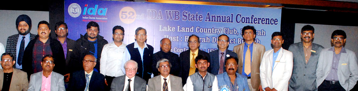
Upcoming Events
1. Article Title.
2. Author Details.
3. Abstract.
4. Keywords.
5. Corresponding Author details.
July 2018
Volume : 34
No.: 2
Dr. Aritri Lahiri,Prof. (Dr.) Shabnam Zahir,Prof. (Dr.) Gautam Kundu,Dr. Paridhee Jalan
Abstract: Child abuse is a very crucial yet ignored topic of discussion in our country. Vast number of children in our country are abused either physically, sexually, emotionally or economically. Dentists and especially pedodontists are in a strategic position to diagnose and report cases of abuse and neglect to the rightful authorities as the oral cavity shows many signs of abuse. As knowledge and awareness among dentists of all specialities regarding abuse and neglect of children is poor, we often fail to fulfil our role by reporting such cases and in punishing the offenders. Increasing the knowledge and training of dentists in this arena will help in protecting children from wrong at the earliest possible stage and thus securing their future.
Dr. Krunal S. Soni,Dr. Satabdi Saha,Dr. Niharika .,Prof. (Dr.) Subrata Saha
Abstract: Ozone therapy has successfully being used in the medical field for treatment of various diseases for more than 100 years. Introduction of ozone therapy has truly revolutionized dentistry. Ozone therapy is completely painless, noninvasive and has advantages of lack of side effects or adverse reactions, increased patient's acceptability and compliance thus making it an ideal treatment option for pediatric patients where patient compliance is of prime importance. This article reviews the clinical application of ozone in pediatric dentistry.
Dr. Krunal S. Soni,Prof. (Dr.) Subrata Saha,Dr. Shubhabrata Pal,Dr. Niharika .,Dr. Satabdi Saha
Abstract: Pharmacological behavior management in pediatric dentistry includes measures involving anxiolysis to minimal sedation to even induction of general anesthesia. It can be used safely with great effect in patients who are unable to receive treatment for various reasons such as young age, compromised state of mind, or health. Use of minimal and moderate sedation is usually more beneficial because of its minimal pre-operative preparation and acceptable comfort level of patients post-operatively as they require no or minimal hospitalization. Credentials of N2O include its simple titration method, superior analgesic, anxiolytic, amnestic properties rapid onset of action, and equally rapid post-operative recovery. It is also preferred over other drugs in inhalation sedation because of its low tissue solubility, minimum alveolar concentration value that accounts for its rapid onset and fast post-operative recovery within minutes and its non-irritant property to the respiratory tract. N2O-O2 inhalation sedation aims at anxiolysis by raising the pain threshold of children during dental treatment with almost negligible adverse events and establishing a healthy communication with them for a cooperative future dental treatment.
Dr. Siddhartha Chatterjee,Dr. Ananya Das,Prof. (Dr.) Sudip Chakraborty
Abstract: The temporomandibular joint (TMJ) is composed of the temporal bone and the mandible, as well as a specialized dense fibrous structure, the articular disk, several ligaments, and numerous associated muscles. Anatomically the TMJ is a diarthrodial joint, which is a discontinuous articulation of two bones permitting freedom of movement that is dictated by associated muscles and limited by ligaments. As part of the stomatognathic system, the TMJ allows simple and complex movements such as free movement during phonation, swallowing, and breathing, as well as different types of load-bearing movements during incision and chewing.The innervation of the TMJ has been studied extensively and includes the auriculotemporal, masseteric, and deep posterior temporal nerves. Of these, the auriculotemporal nerve, because of its course, is at the greatest risk for irritation or entrapment. The knowledge of the course and anatomical relation of facial nerve and its temporal branches is fundamental for successful temporomandibular joint (TMJ) surgery since these branches are susceptible to damage during this procedure. The risk of lesioning the facial nerve in this type of incision is approximately 5%. The proximal portion of the maxillary artery and the masseteric artery is in close relation with the subcondylar region of the mandible. The maxillary artery has a variable course which can be further distorted by the fibrotic changes or ossification process in the TMJ region in temporomandibular joint ankylosis. In this review article we have tried to discuss the surgical anatomy for a successful TMJ surgery.
Dr. Ashit Kr. Pal,Dr. Somen Bagchi,Dr. Parthasarathi Biswas,Dr. Zenab Khan,Dr. Parthasarathi Saha
Abstract: Gingival hyperpigmentation is a major concern for many people for esthetics. It is corrected by various periodontal plastic surgical procedures. Various techniques had been introduced like scalpel, electrosurgery, cryosurgery, laser as well as power driven rotary abrasive method and masking procedures like FMG and ADM allografts. This paper will highlight a comparative evaluation between scalpel and rotary abrasive technique.
Dr. Parvin Sultana,Dr. Shankhanil Dev,Dr. Subhamay Chakraborty,Prof. (Dr.) Gautam Kundu,Dr. Sudipta Kar
Abstract: Young children are very much aware of their appearance in 21st century. Children are becoming very conscious about their esthetics, because they live very much in an era of peer influence. Stainless steel crown (SSC) is a very useful treatment in child patients. They are mainly used following the endodontic treatment in deciduous molar. Stainless steel crowns have shown significant clinical success but its main drawback is unesthetic appearance. This is a case report of a child patient with Stainless steel crown, came to the department of Pedodontics and preventive dentistry with his mother after complete endodontic treatment and stainless steel crown in lower primary second molar. The tooth was clinically asymptomatic, but patient was not satisfied because the visibility of Stainless steel crown during smile had become embarrassing in front of his friends. So to satisfy the child, Stainless steel crown has been modified by chairside veneering to improve the esthetics and restore his smile.
Dr. Somen Bagchi,Dr. Tirthankar Debnath,Dr. Ashit Kr. Pal,Dr. Zenab Khan,Dr. Parthasarathi Saha,Dr. Parthasarathi Biswas
Abstract: Pleasant smile is considered a symbol of beauty and well being which extensively deals with soft tissue harmony. Gingival fibromatosis is manifested as a benign enlargement of the gingiva involving both the arches, hence causing major aesthetic, phonetic and masticatory problems. Diode LASERs are used for the soft-tissue manipulations like gingivectomy, gingivoplasty, operculectomy or frenectomy because of its highly absorbable property by melanin and hemoglobin, accompanied by improved epithelization and wound healing, hence, overcoming bleeding, trauma, pain, postoperative edema and overall patient discomfort. In this case report, split mouth gingivectomy was done by 940 nm diode LASER and conventional scalpel method, for the management of gingival overgrowth and a comparative evaluation done with different parameters. With all limitations, it can be concluded that, although both the techniques were equally effective for surgical removal of gingiva, but, initially post operative healing, aeasthetics and patient satisfaction is more in gingivectomy by LASER along with less pain, than in traditional scalpel technique. Whereas, scalpel method shows better result in the long run.
Dr. Chiranjan Guha,Dr. Ipsita Maity,Dr. Paromita Mazumdar
Abstract: Perforations represent pathologic or iatrogenic communications between the root canal space and the attachment apparatus. The causes of perforations are resorptive defects, caries, or iatrogenic events that occur during and after endodontic treatment. Case report: A 27-year-old male patient reported to the Department. Clinical examination revealed that access preparation had been attempted in the grossly carious maxillary right central incisor the access opening was carefully examined and no bleeding was present. An intraoral periapical radiograph was taken with a #25 size K file placed in the perforation, The lateral walls of the perforation were refined and cleaned. A #25 size K file was used to establish the length from the incisal reference point up to the perforation site inside the root canal. Working length was established. GuttaFlow Bioseal was mixed and carried to the perforation site with the help of lentulo spiral and packed with an appropriately fitted plugger. Repair of the perforation was carried out by placement of the sealer. Cleaning and shaping was done upto file size F5. Canal was irrigated with 1.5 mL of 2.5% NaOCl. The root canal was obturated with the same sealer such that the sealer was allowed to flow out of the access cavity. Conclusion: advances in technologies have also provided for more controllable and better treatment outcomes, either surgically or nonsurgically.
Dr. Parthasarathi Saha,Dr. Parthasarathi Biswas,Dr. Zenab Khan,Dr. Somen Bagchi,Dr. Ashit Kr. Pal
Abstract: Excessive exposure of gingival tissue during smile has been a matter of aesthetic embarrassment for many patients. Excessive gingival display may not be present with any pathologic condition but it definitely affects the patient's psychosocial behaviour. This case report will highlight a surgical procedure to restrict hyperactive upper lip and a mild vertical maxillary excess by removing a strips of mucosa with the help of partial thickness from maxillary buccal vestibule on both the sides and suturing the lip mucosa with mucogingival junction, thereby reducing gingival display.